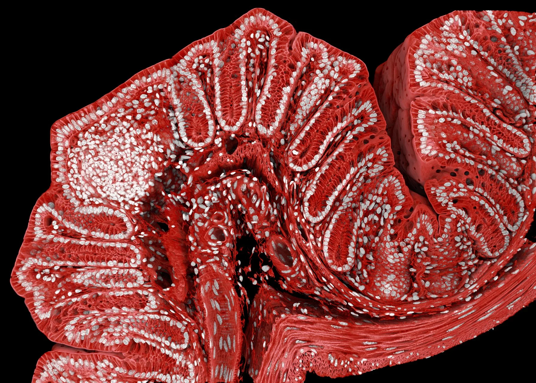
Marius made place 18 in the Nikon Small World Photomicrography Competition, a reference competition for light microscopy images, with his mouse colon image. The 3D rendering shows a 300 um thick cleared section of a mouse colon, where nuclei were stained with DAPI and the cell membranes were tagged endogenously with GFP. The volume was imaged with a Stellaris 8 and a 20x objective over several days at FMI and reconstructed with Drishti, an open source software common in the macroscopic imaging (MRI / CT) field. We use this image and others like it as introductory and teaching material.