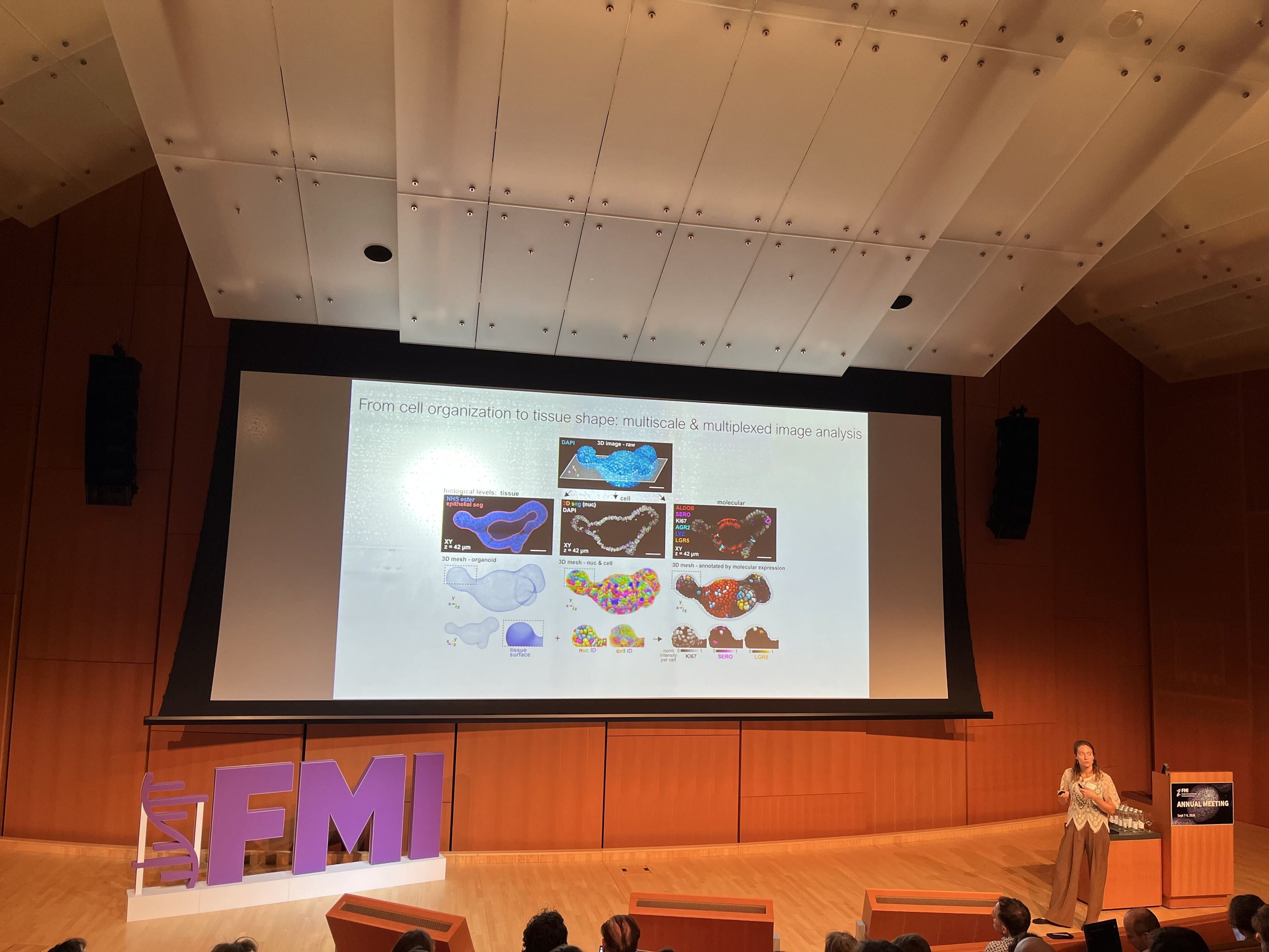
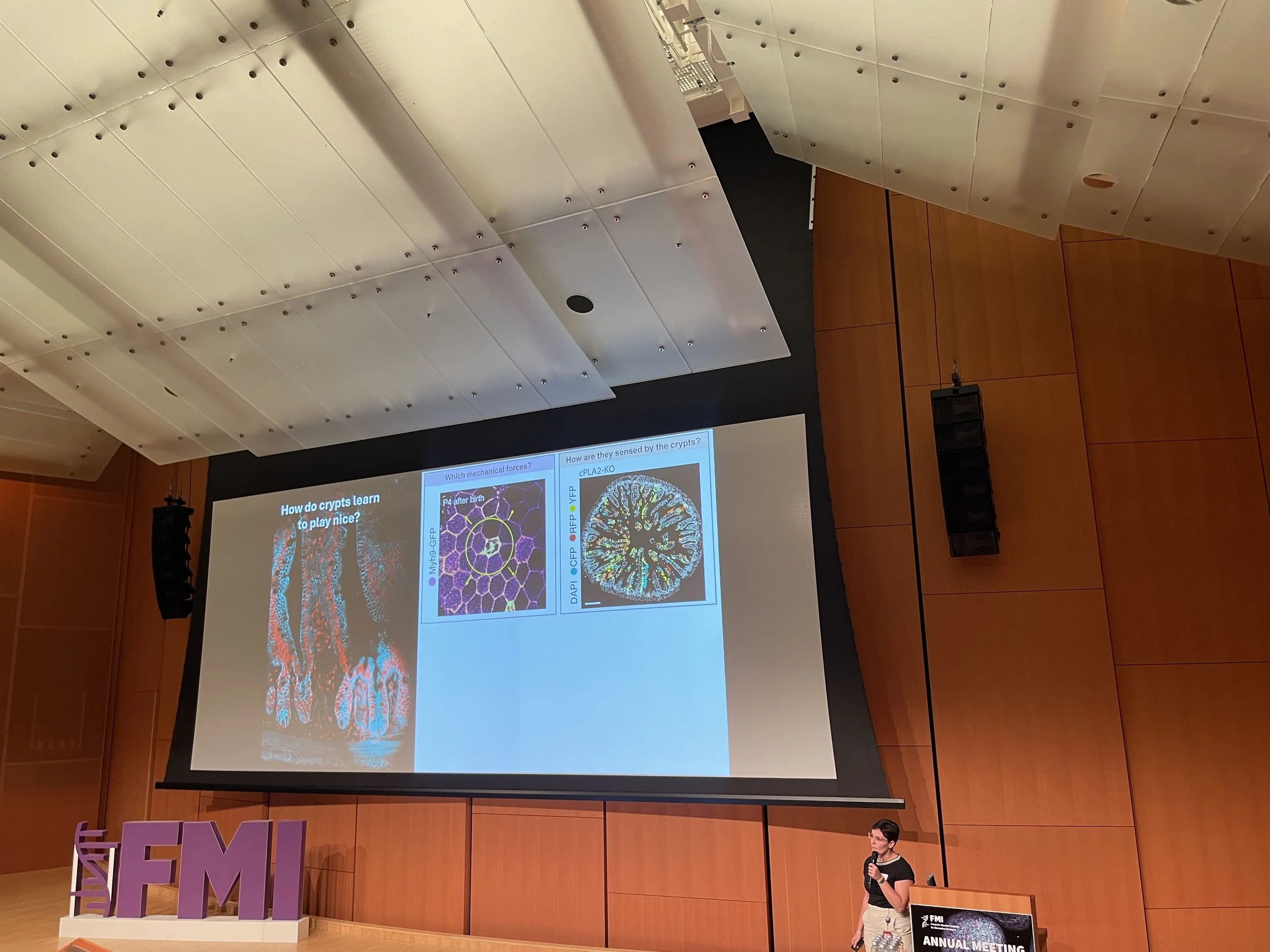
Our paper is out in Cell!

We are thrilled to share that our lab's latest research has been published in Cell.

Check it out here: Multiscale integration of tissue and chromatin context converts cell heterogeneity into stable intestinal patterning: Cell

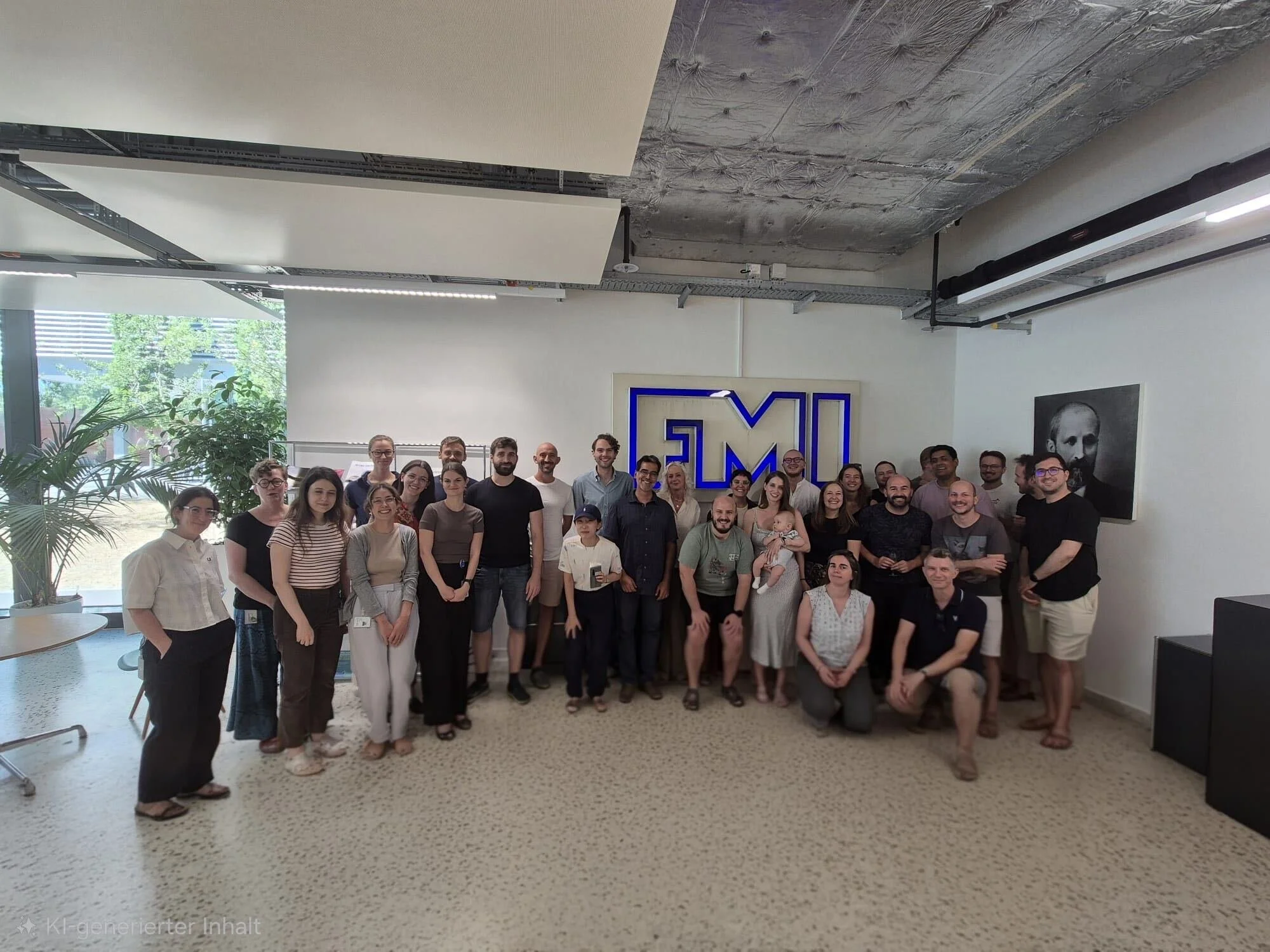
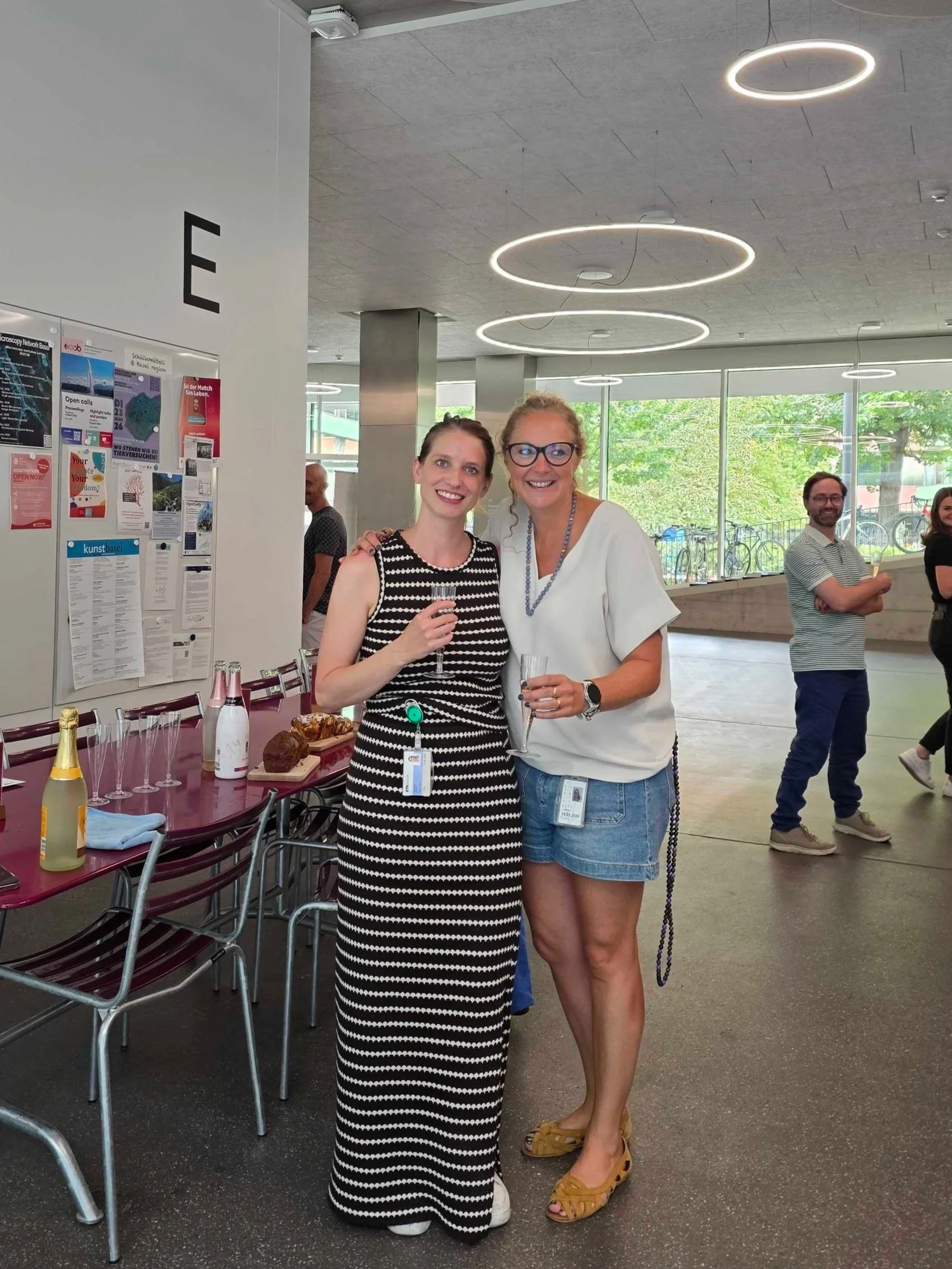
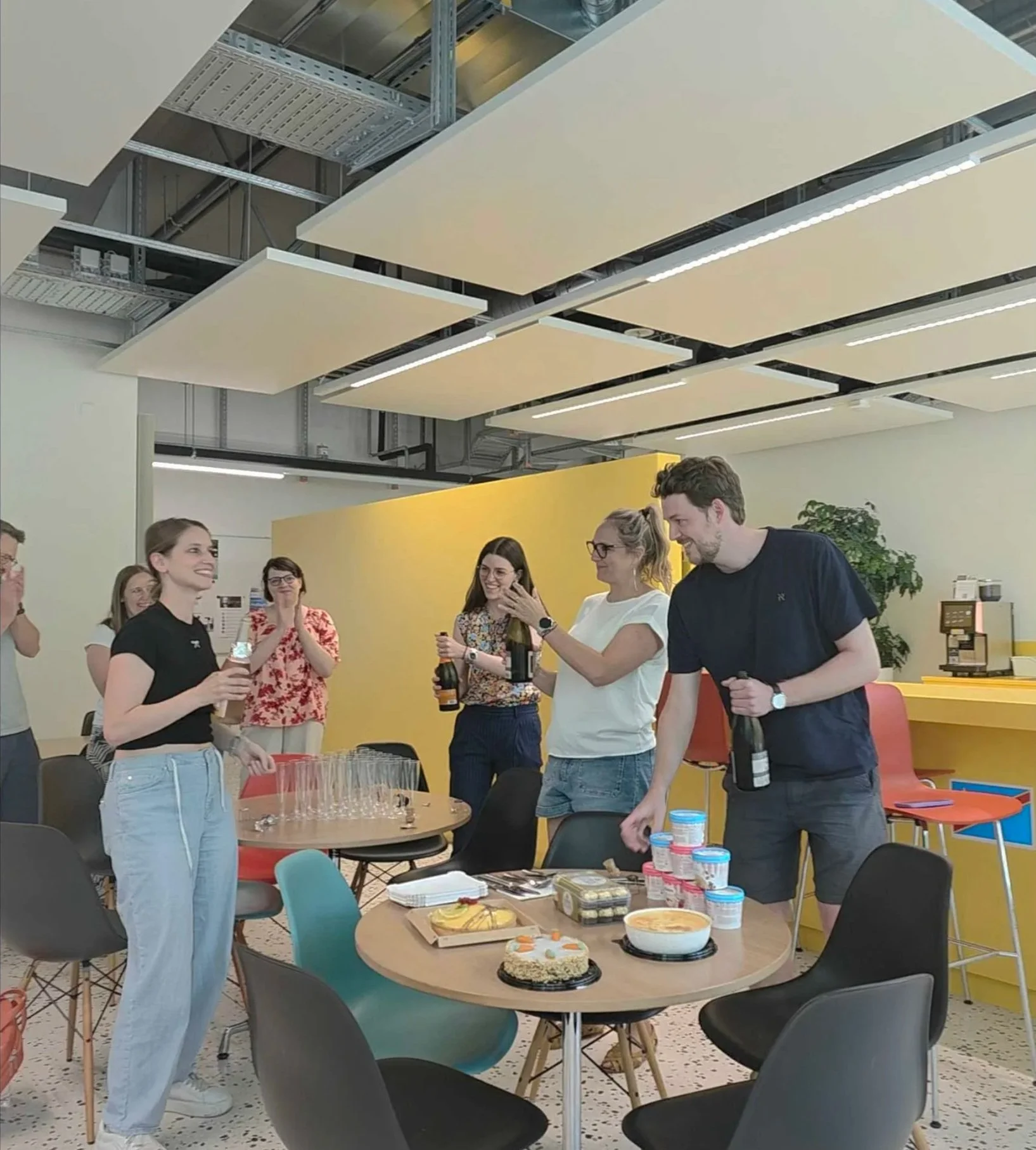
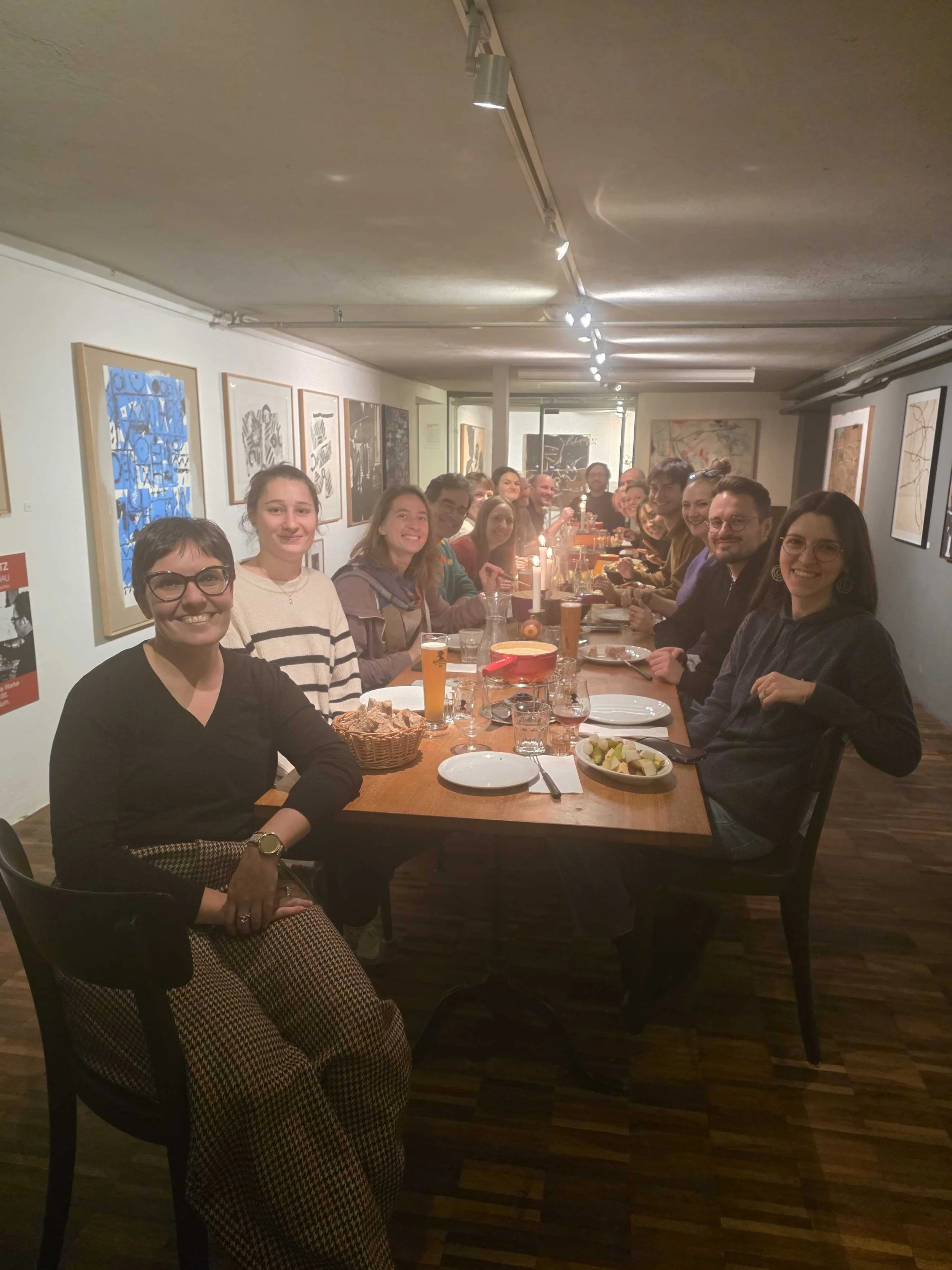
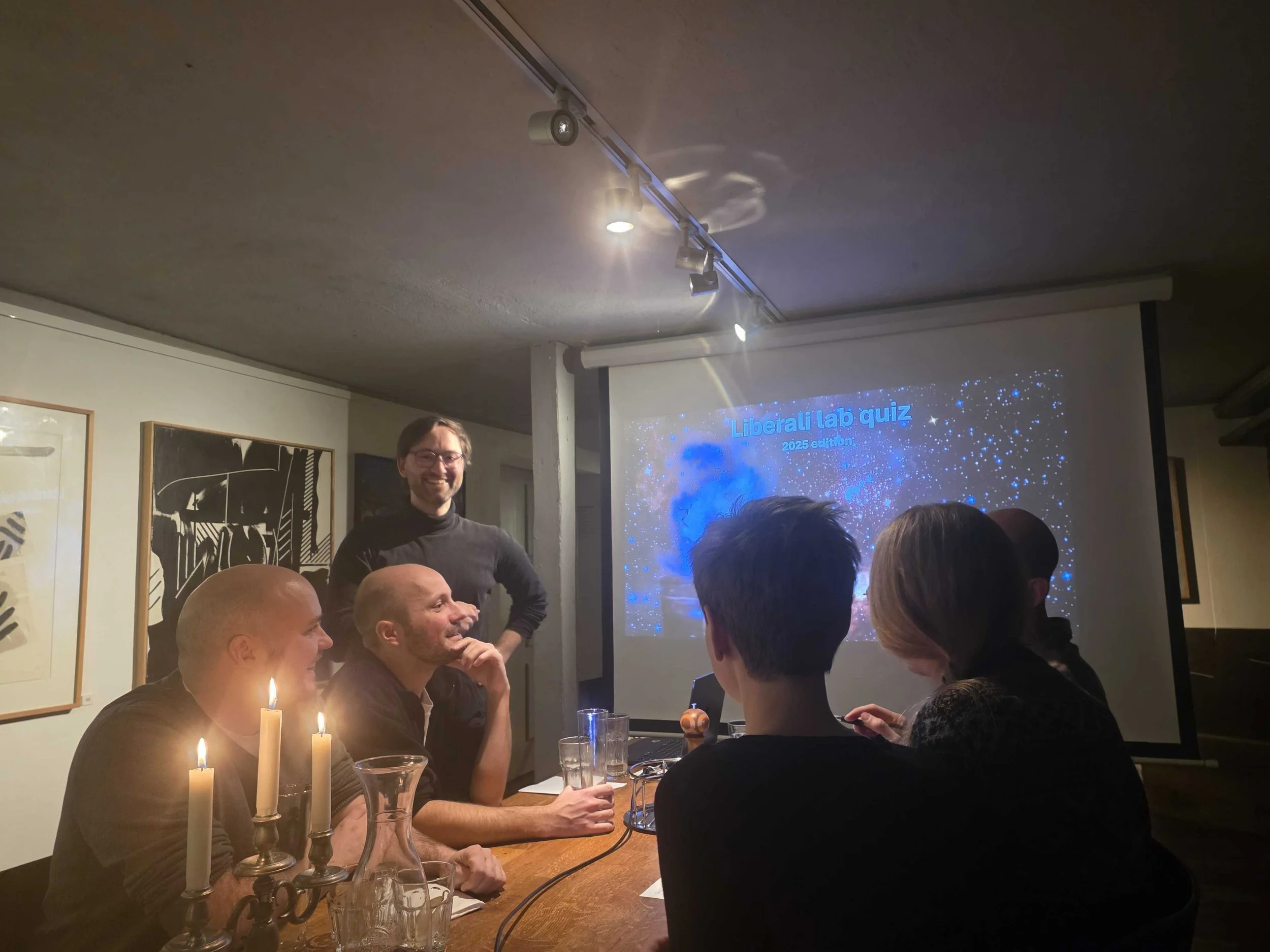
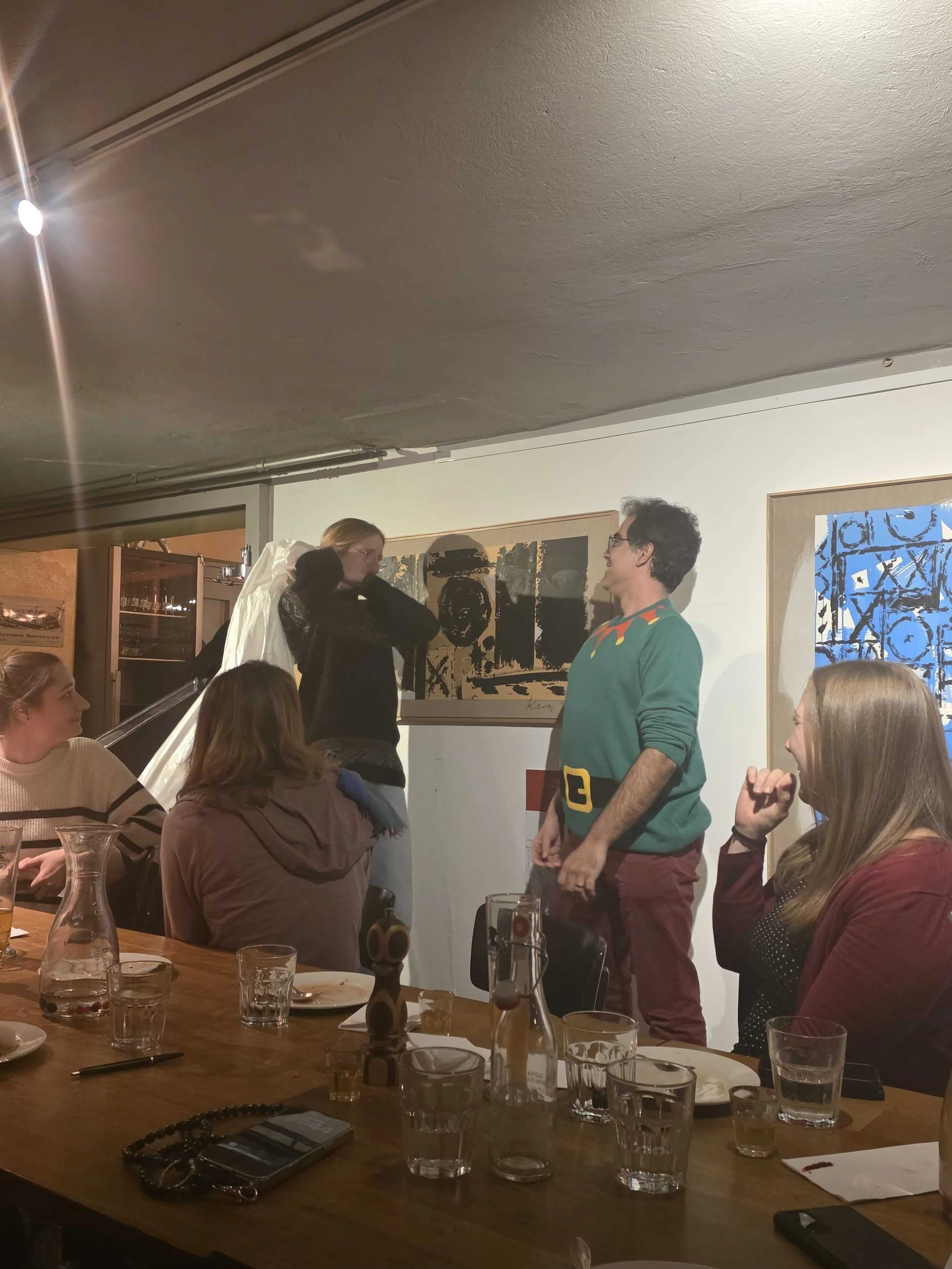
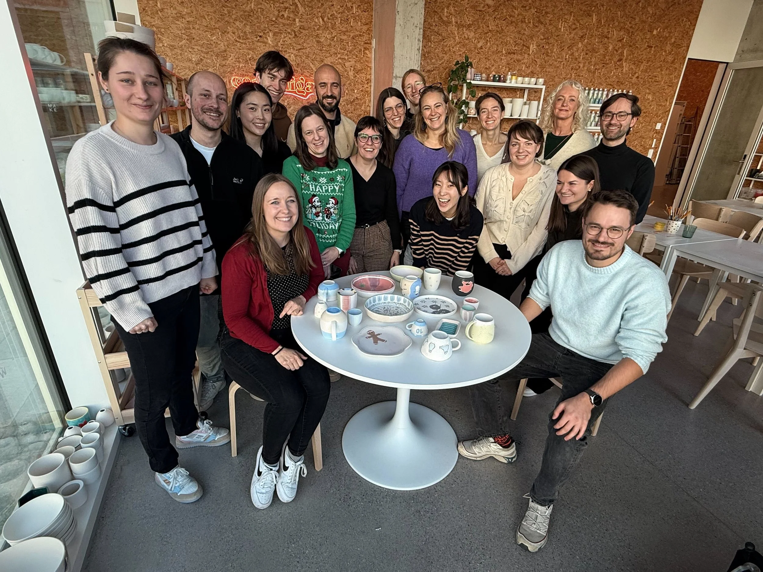
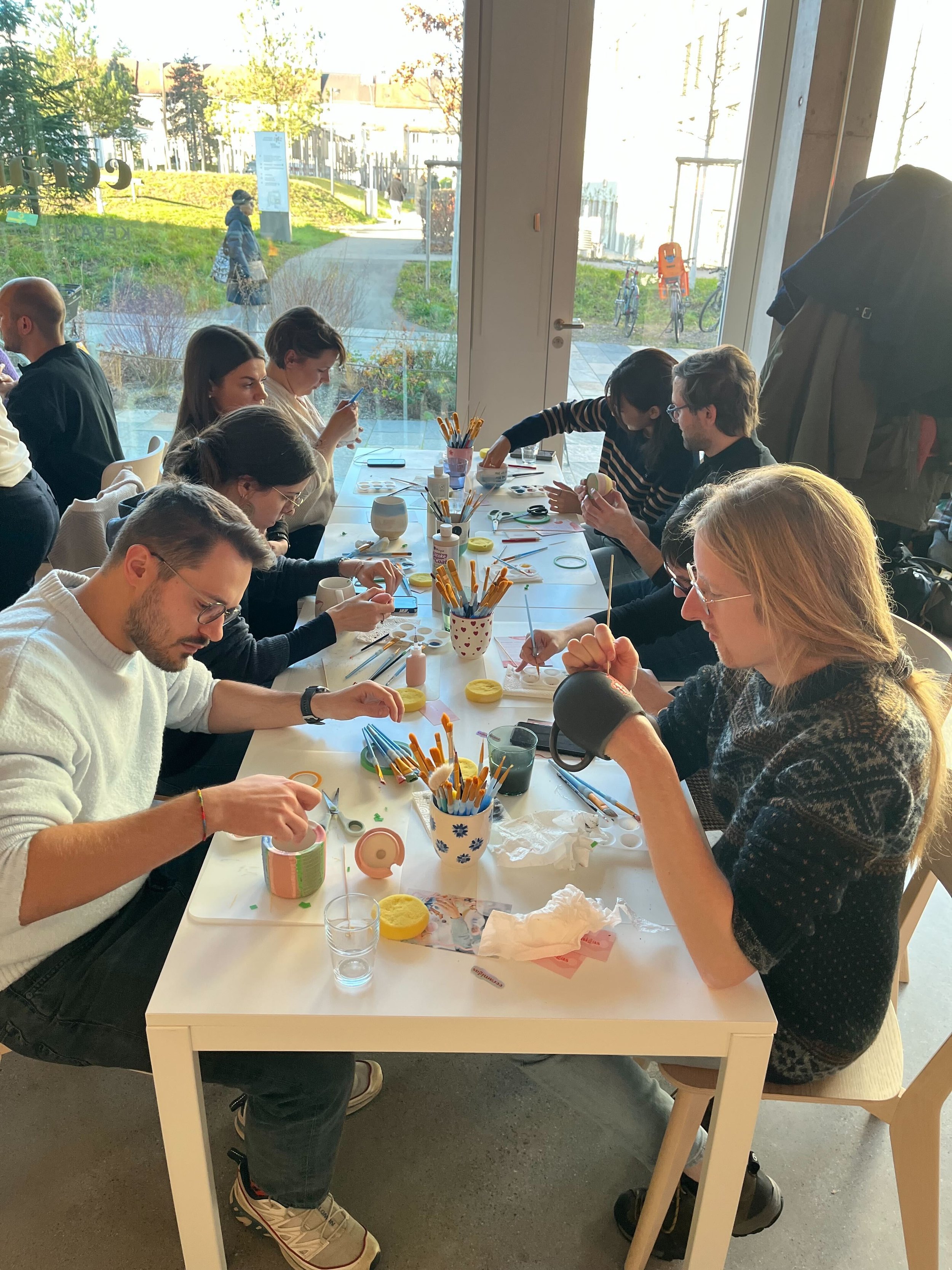
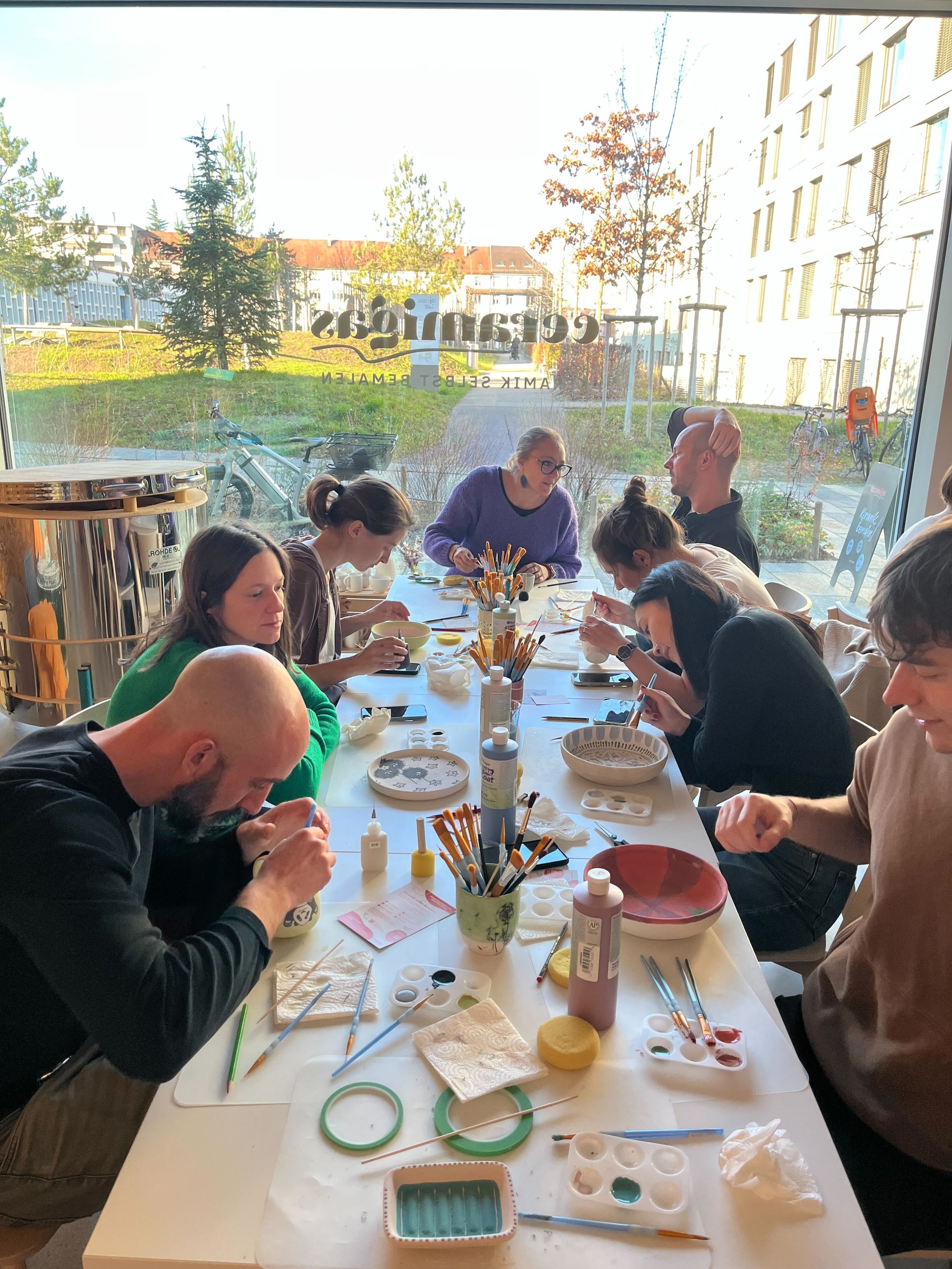
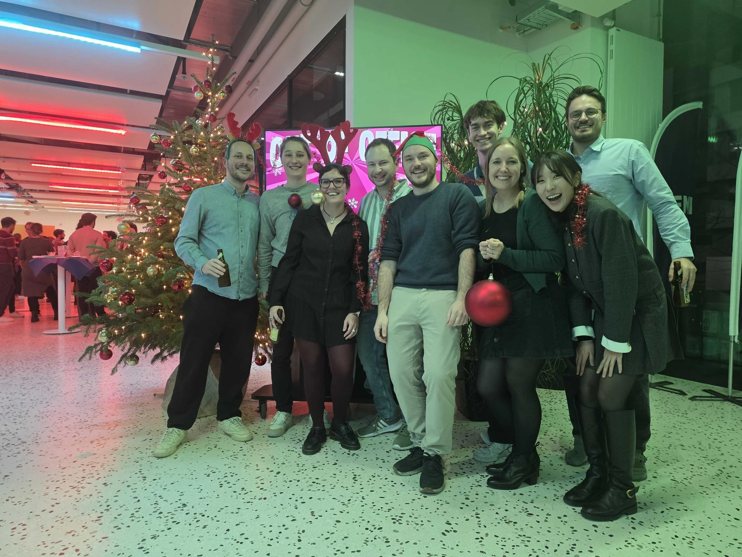
This work represents years of dedication and collaboration, and we are incredibly proud of what the team has accomplished. To celebrate this milestone, we gathered for a small party with the paper's authors, marking the occasion with well-deserved cheers. Congratulations to everyone involved!!

Public lab tour

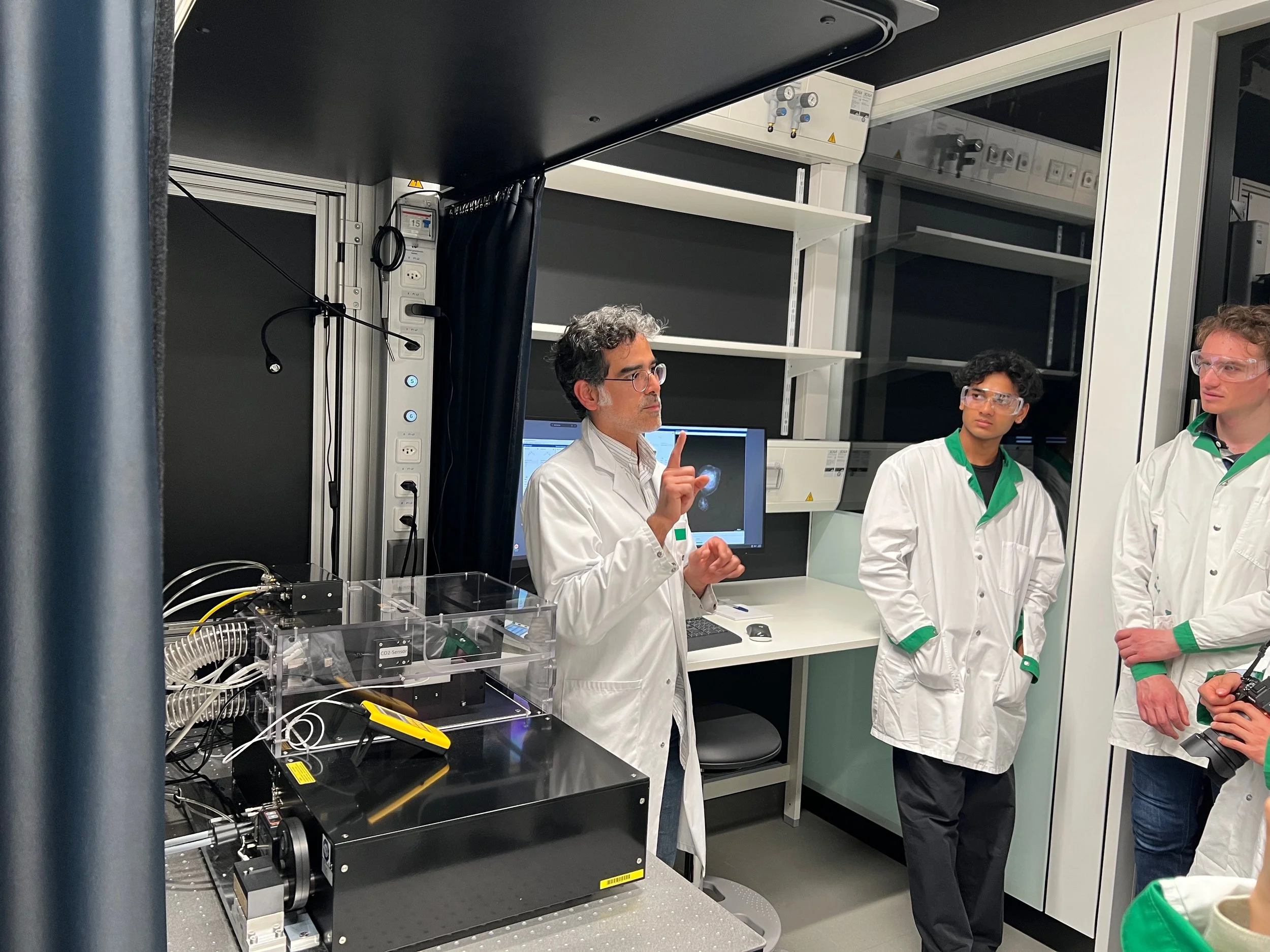
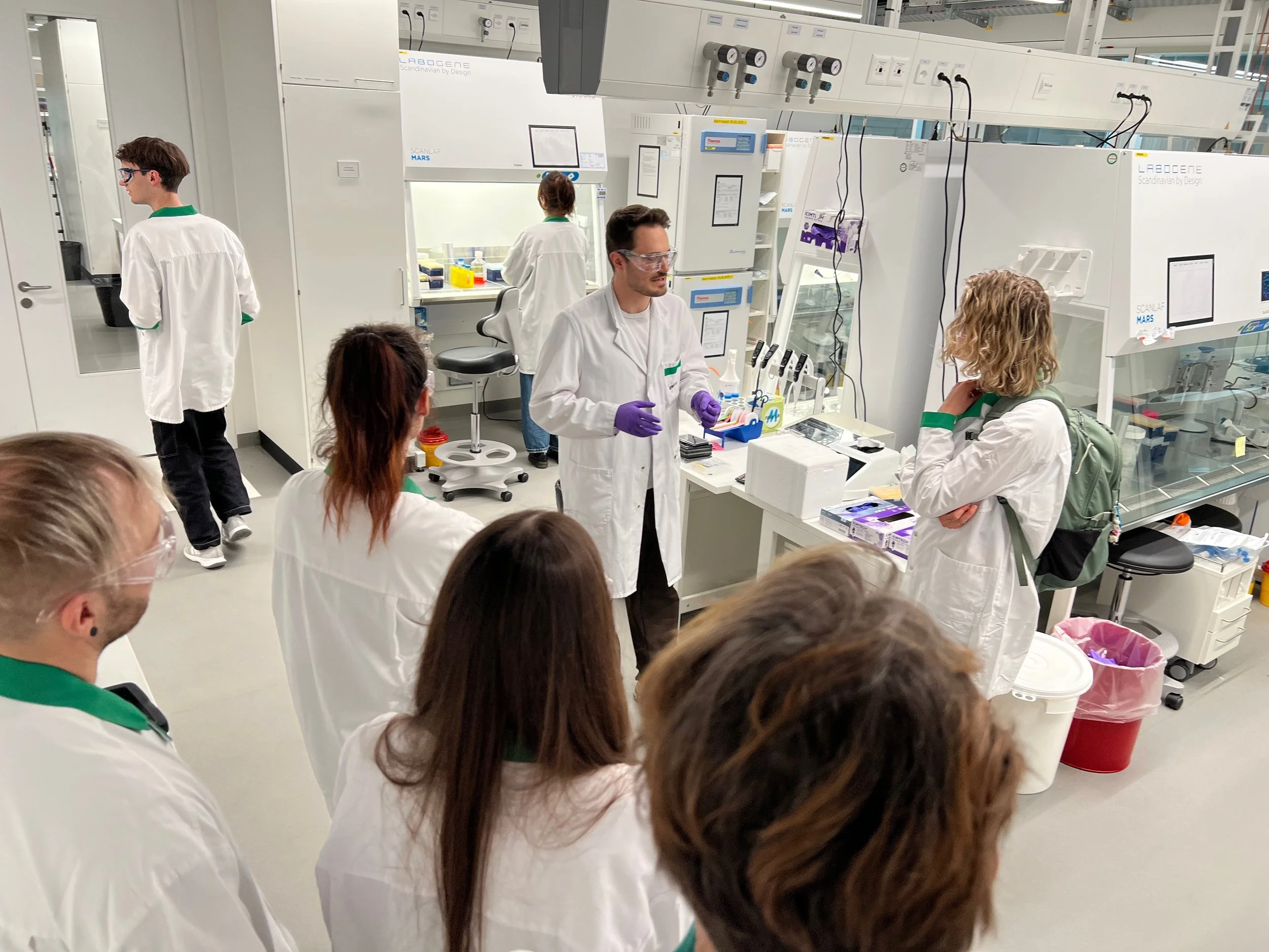
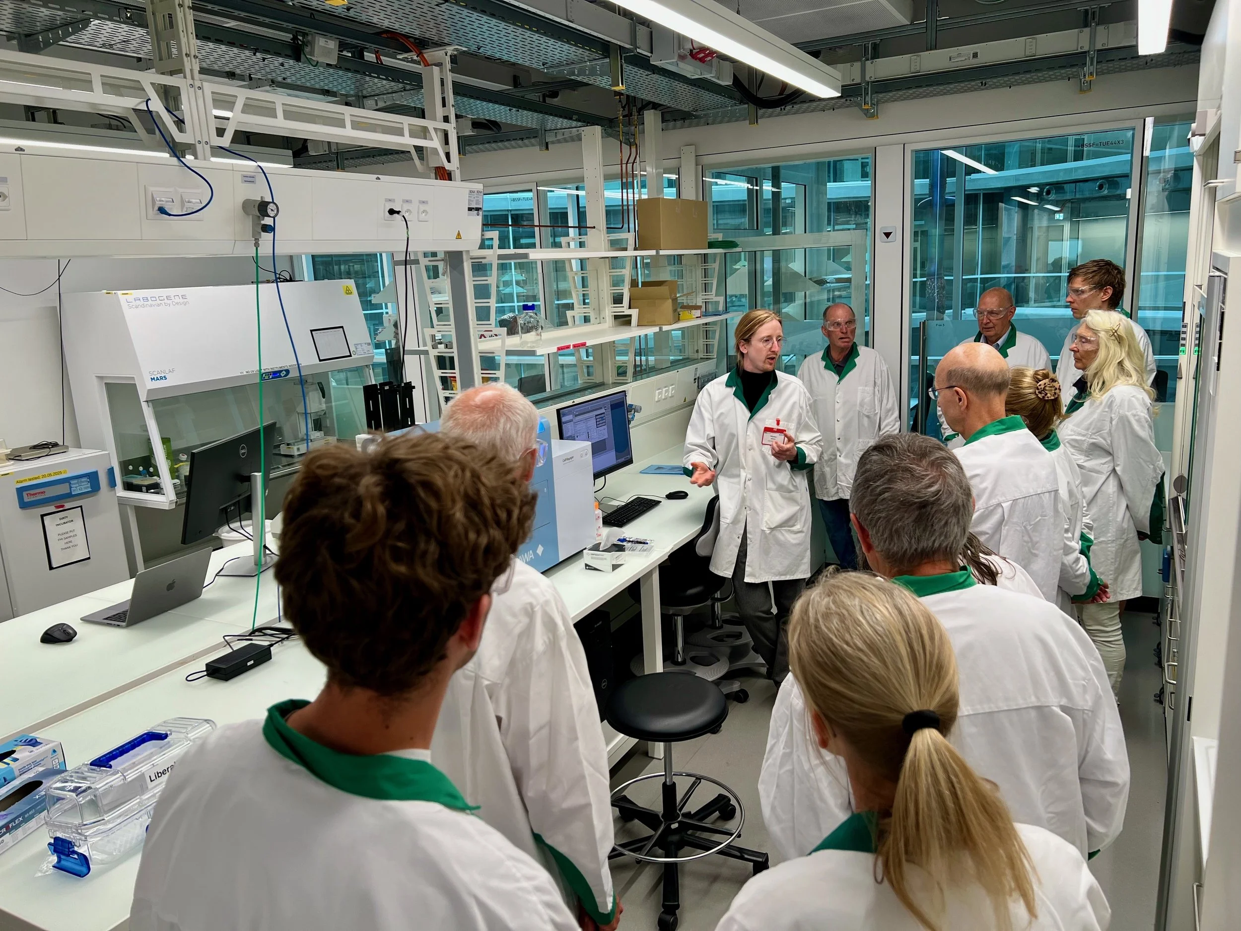
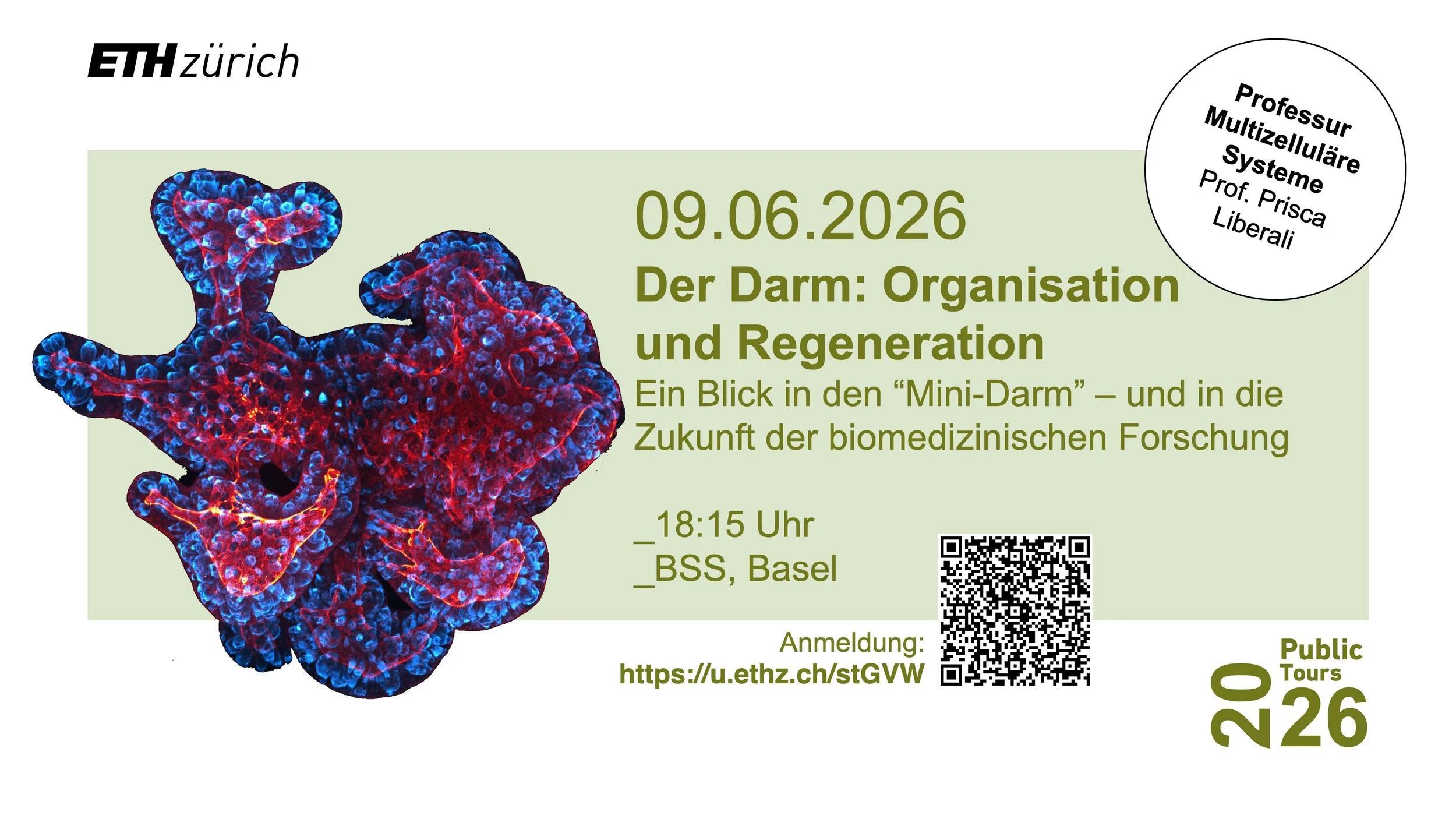
Our lab recently hosted a public lab tour that was open to everyone. From the high-content screening facility to the light-sheet microscopy room, guests were able to discover the technologies and approaches that drive our scientific work. It was a wonderful opportunity to engage with the public, share our research, and inspire curiosity about science!

Flavours of DBSSE event

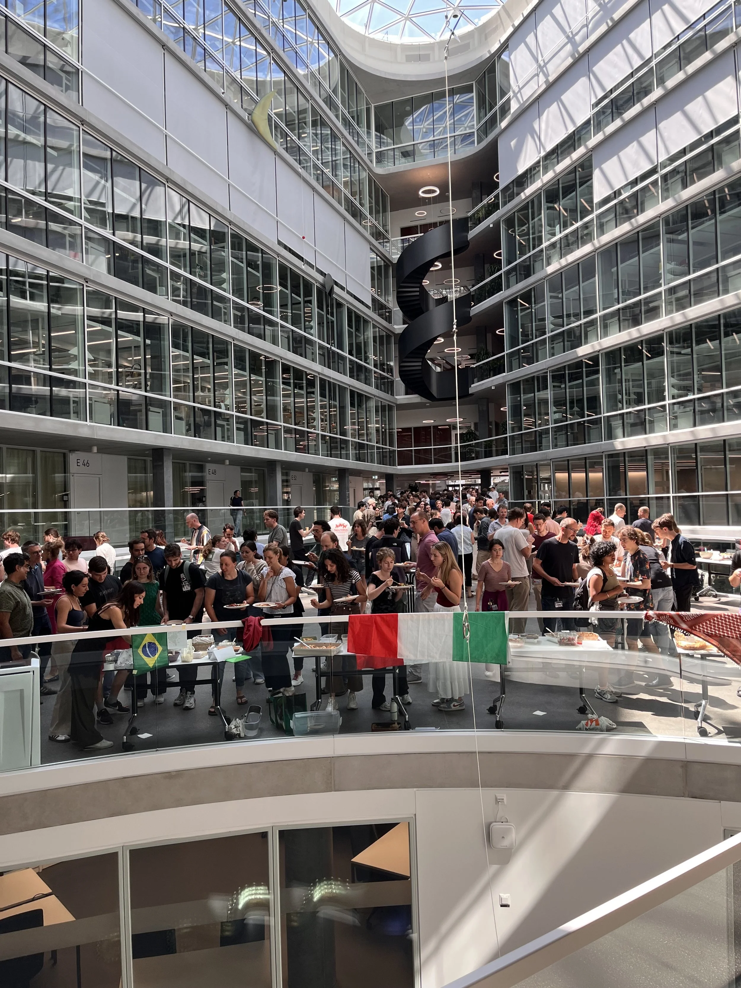
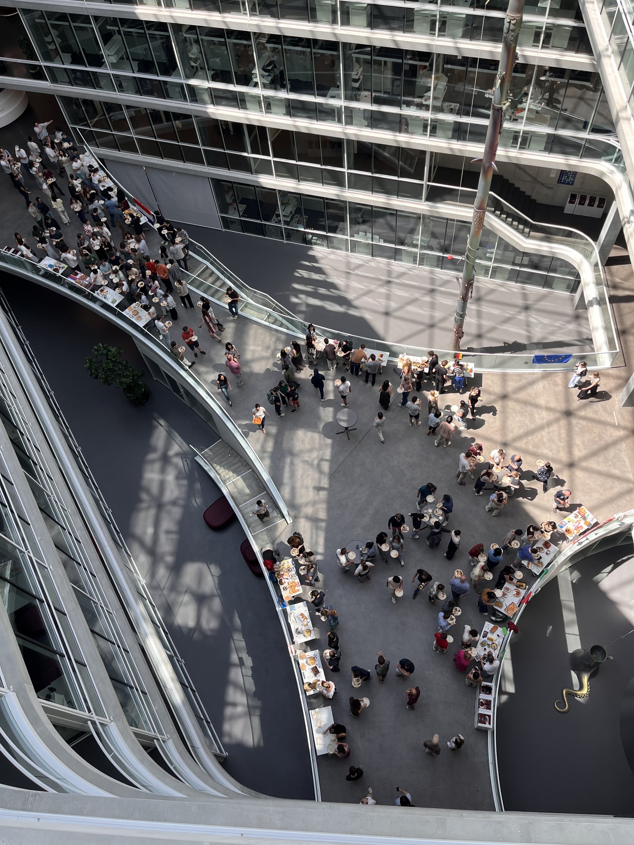
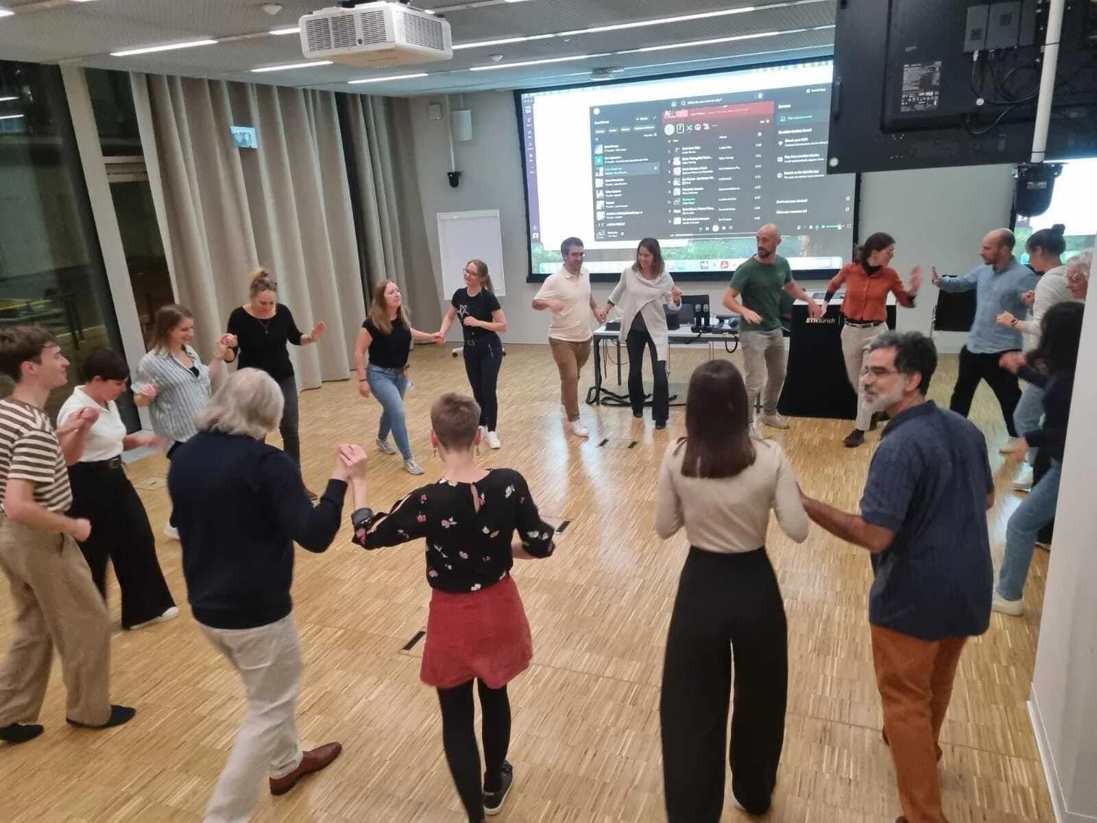
Our lab recently organized "Flavours of DBSSE", a culinary event that brought the whole department together in the most delicious way possible. The idea was simple: set up tables representing different countries and regions, each filled with homemade dishes reflecting the rich cultural diversity of our department.

It was a wonderful success and it reminded us how many cultures, backgrounds, and traditions make DBSSE the unique place it is. :)

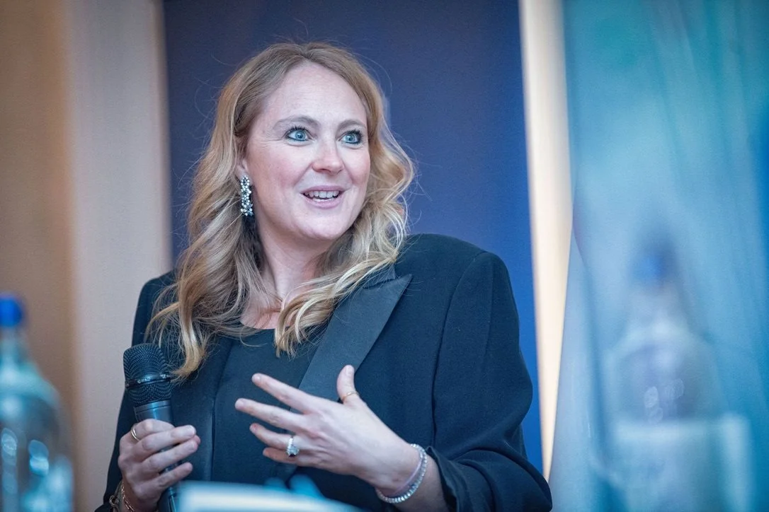
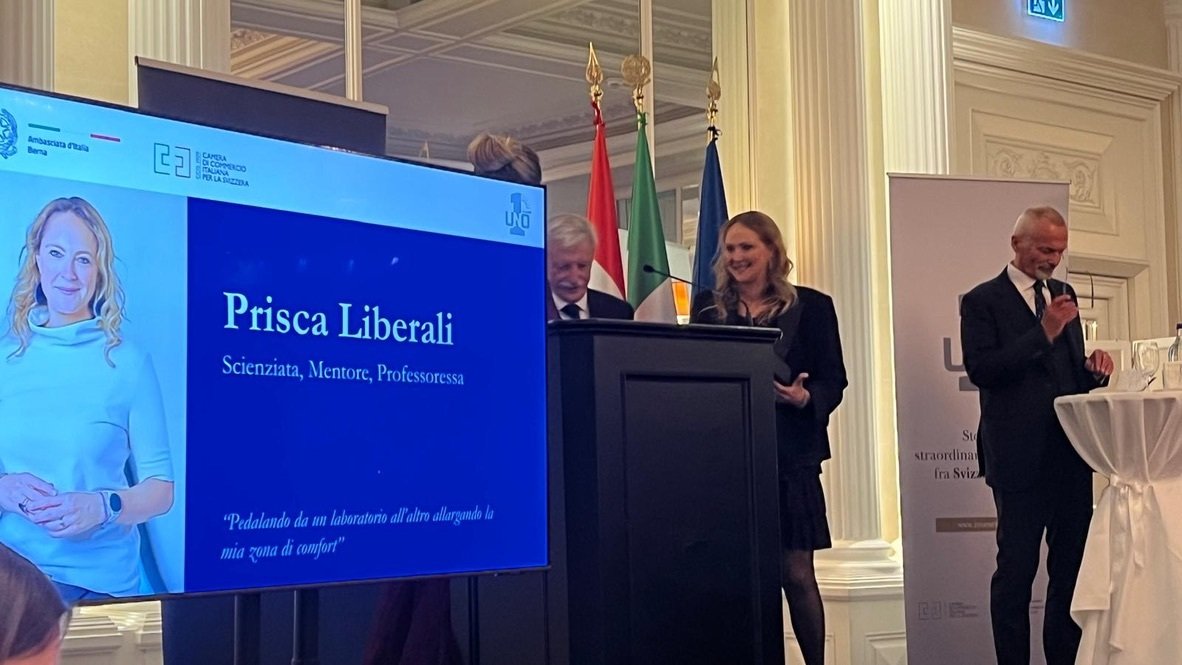
Prisca Receives I Numeri UNO Award

Prisca has been honored with the Il Premio I Numeri UNO for “Storie di Straordinaria quotidianità fra Svizzera e Italia.” This prestigious award recognizes women and men who, across diverse fields and career paths, have distinguished themselves through their contributions to strengthening positive relationships and fostering mutual respect between Italy and Switzerland.

We are so happy for you, Prisca. Congratulations on this well-deserved recognition!

Marius' colon image in the Nikon Small World Top 20

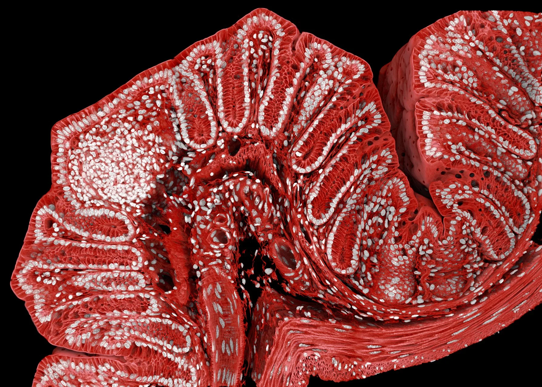
Marius made place 18 in the Nikon Small World Photomicrography Competition, a reference competition for light microscopy images, with his mouse colon image. The 3D rendering shows a 300 um thick cleared section of a mouse colon, where nuclei were stained with DAPI and the cell membranes were tagged endogenously with GFP. The volume was imaged with a Stellaris 8 and a 20x objective over several days at FMI and reconstructed with Drishti, an open source software common in the macroscopic imaging (MRI / CT) field. We use this image and others like it as introductory and teaching material.

The bell of success

During our 10th year celebration, Prisca rang our bell and with that our new lab tradition was born: the “Bell of Success”.

From now on, every time someone in the lab achieves something, whether big or small, they will ring the bell. When the bell rings, we pause our work to come together and celebrate the moment all together.

Looking forward to our fun moments through sharing our moment of success!